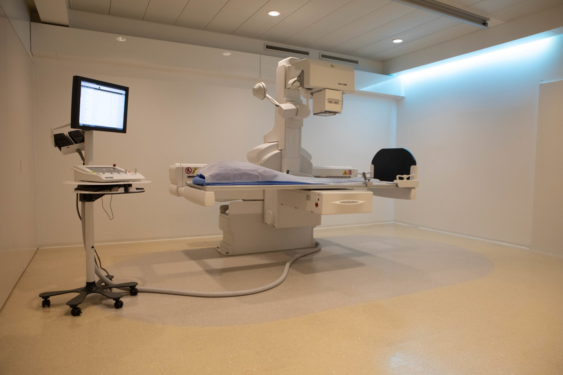
Computer tomography (CT)

Scanning is a diagnostic imaging technique that uses a beam of X-rays to ‘scan’ a given area of the body to create cross-sectional images. The technique involves measuring the absorption of X-rays by the tissue, with the rays being absorbed to a greater or lesser extent depending on the density of the tissue. The data collected is then analysed by a computer, which reconstructs the images. Unlike X-rays, which focus more precisely on bones, CT scans can be used to view most organs, even through bones. Different anatomical structures absorb X-rays in different ways.
- CT scans of adults and children
- Cardiovascular imaging (angio-scan and coro-scan)
- Digestive imaging (virtual colonoscopy, entero-CT, abdominal CT)
- Neurological and spinal imaging
- Osteoarticular imaging (including arthro-scanner)
- Lung and thoracic imaging (low-dose or injected CT))
- Urogenital imaging (low-dose scanner and uro-scanner)
- Oncology imaging
- Dentascan
- Virtual colonoscopy
- Cervical infiltration
- Lumbar infiltration
There are no contraindications to having a CT scan. However, certain physiological conditions (pregnancy or risk of pregnancy, breast-feeding) or pathological conditions (diabetes, renal insufficiency, allergy) require special precautions.
A CT scan is a fast procedure, but depending on the type of examination to be carried out, the entire procedure, including preparation, positioning and scanning, may take up to 30 minutes (or even 45 minutes). For abdominal, thoraco-abdominal or brain scans: fast at least 3 hours before the examination. For virtual colonoscopies, a preparation, protocol and explanations will be sent to you a few days before the examination.
The examination form signed by your doctor.
Any previous examination results.
Your health insurance card or, in the event of an accident, your declaration.
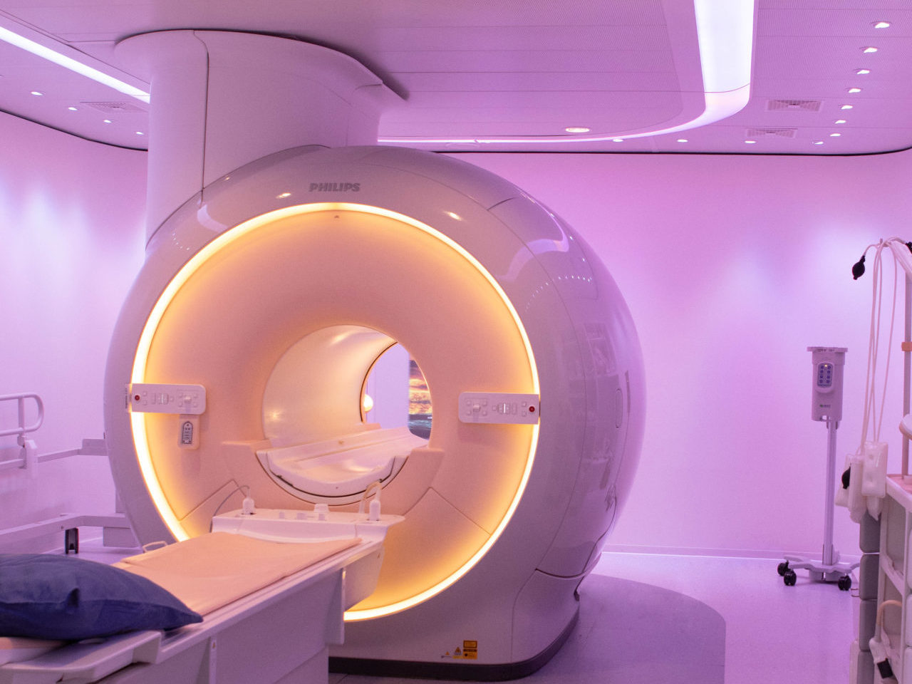
You will lie on a bed that moves in a wide ring, usually on your back and alone in the examination room; we will be able to communicate with you via a microphone. The team will be right next to you, behind glass. They can see and hear you throughout the examination. If necessary, they can intervene at any time. Depending on the area being examined, your arms will be at your side or behind your head. The examination is generally quick. Your cooperation is important: you should try to remain still; in some cases, we will use the microphone to tell you when to stop breathing for a few seconds. You will stay in the scanner room for an average of 15 minutes. Depending on the case, some examinations require an intravenous injection, usually in the crease of the elbow, a drink or an enema.
Spectral X-ray scanner. Spectral detector technology simultaneously identifies both high-energy and low-energy photons, enabling you to visualise both anatomy and tissue composition. The spectral scanner is dual-energy, so the spectral function is always activated, unlike a conventional scanner.
