Mammography
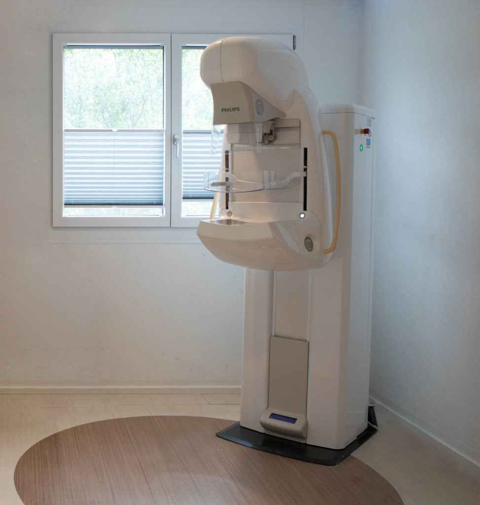
Mammography is an X-ray of the breasts using low-dose X-rays. It enables breast tissue to be analysed, with the aim of detecting any abnormalities in breast tissue and glands.
Compression is achieved using a compression pad specially designed for this type of investigation and adjusted so that it is tolerable for the patient. This compression allows the breast tissue to spread out, making it easier to visualise the breast structures and detect any nodules by increasing the contrast of the image obtained.
Often, at your doctor’s request, the radiologist will supplement the diagnostic mammogram with an ultrasound scan.
It should be noted that from the age of 50, depending on the canton, women are invited to undergo an organised screening mammogram as part of the breast cancer screening programme.
Individual mammography, mammography and ultrasound +/- puncture – biopsy. Canton screening mammography.
If you notice any irritation of the skin under the breast, we recommend that you postpone your examination.
If you are pregnant or likely to become pregnant.
No preparation is necessary. However, on the day of the examination, please refrain from using cosmetics (cream, deodorant, etc.) which could cause false images to appear and hamper the diagnosis.
If you are still menstruating, it is best to book an appointment within ten days of the start of your period, as your breasts will be less sensitive during the examination.
The examination form signed by your doctor or the Canton questionnaire (if screening mammography).
Any results of previous examinations.
Your health insurance card
The examination is carried out standing up, with the patient bare-chested. Your breasts are placed one after the other between two plates. These plates tighten and compress the breast for a few seconds. This examination may be uncomfortable and slightly painful, but it poses no health risk.
Allow 30 minutes for the procedure.
Microdose SI L50 Philips.
