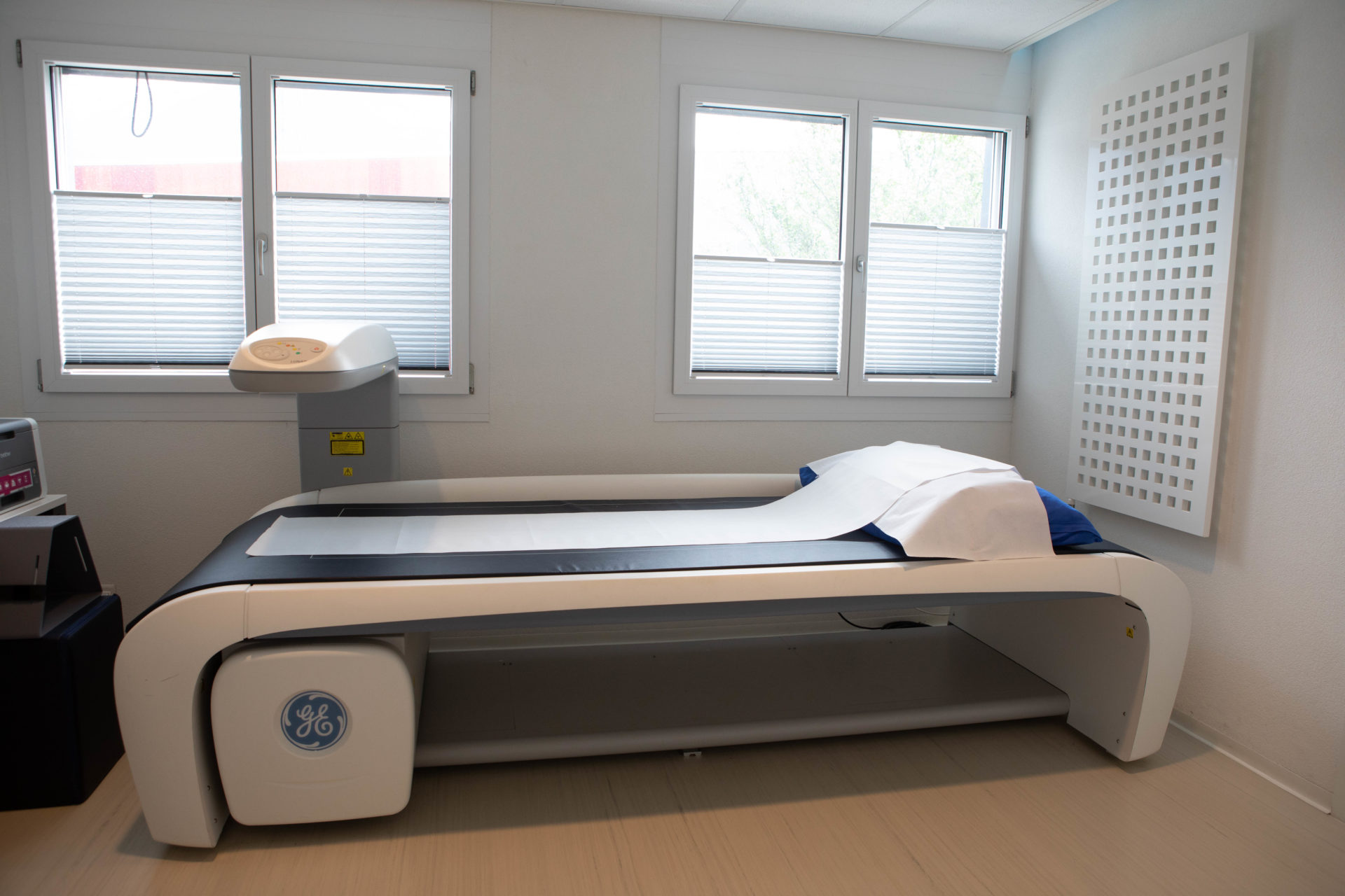
Radiography
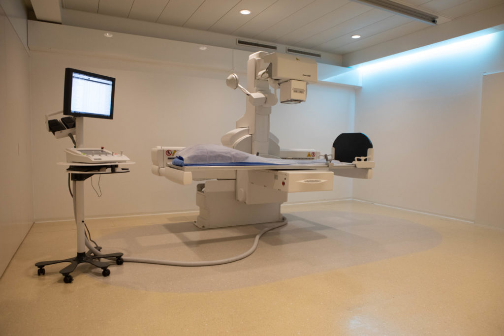
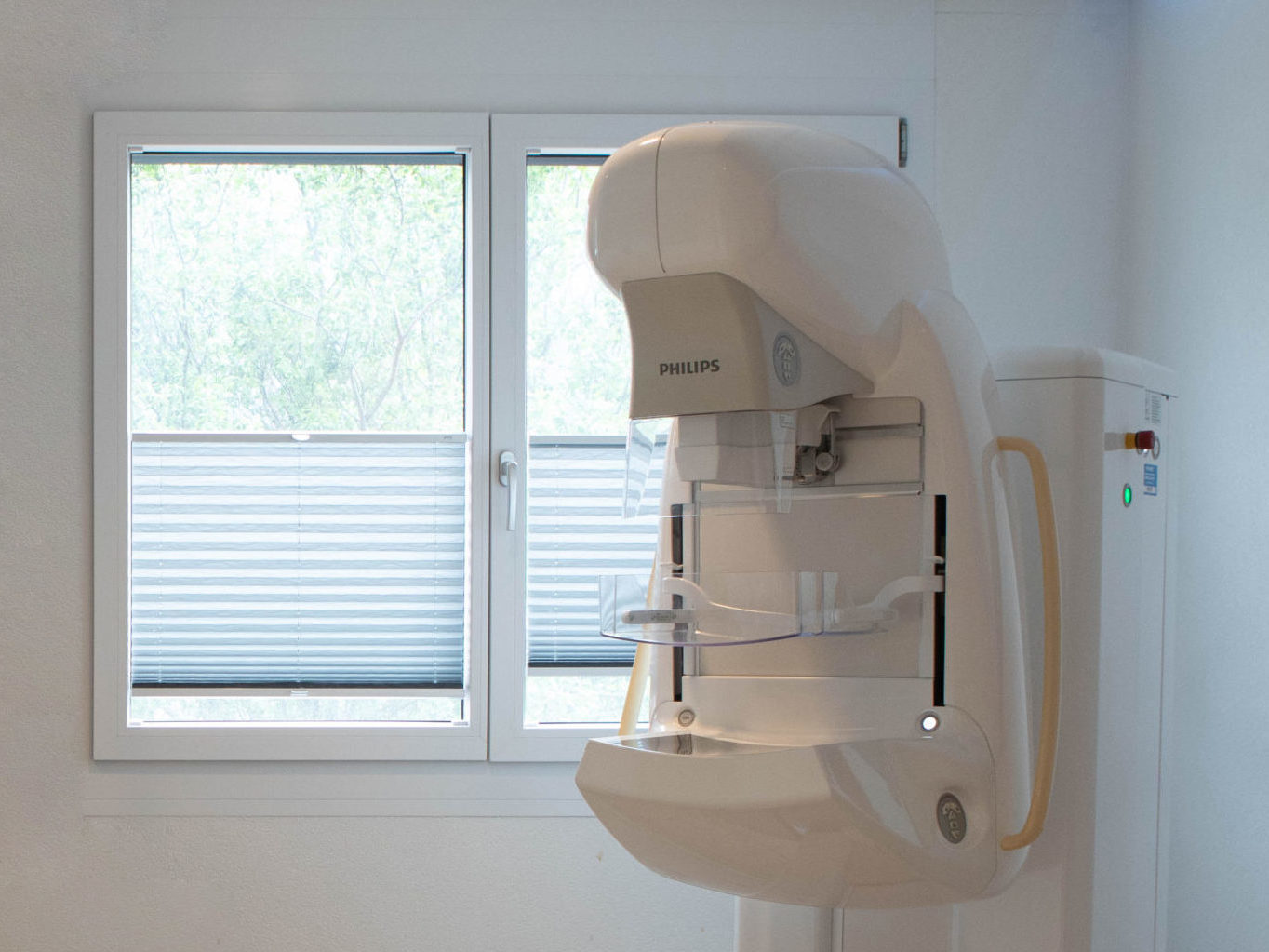
An X-ray is a medical imaging test used to visualise all or part of an area of the body. This examination requires the use of X-rays because of their ability to penetrate tissues to a greater or lesser extent depending on their density. An X-ray source is placed in front of the part of the body to be studied, while a detector is located behind it. The luminous molecules emitted pass through the body, being absorbed to a greater or lesser extent by the tissues in their path. This is how bones and muscles can be distinguished on X-ray images.
- Pulmonary and thoracic
- Neurological imaging
- Osteoarticular imaging
- Spinal column imaging
- Abdomen
If you are pregnant or likely to become pregnant.
Allow around 30 minutes.
You stand, lie down or sit, as appropriate, next to the X-ray machine.
The medical X-ray technician will ask you to adopt various positions, some of which may be uncomfortable, but never painful. Several images are often required to view the organ under examination from different angles (front, profile, etc.). X-rays of both limbs may also be useful for comparison, particularly for children.
Juno Philips
